Upper Leg Tendon Anatomy : Conceptual 3d Gracilis Human Upper Leg Stock Illustration ... : The longissimus (red, in the image above) are located between spinalis and the iliocostalis muscles.
byAdmin-
0
Upper Leg Tendon Anatomy : Conceptual 3d Gracilis Human Upper Leg Stock Illustration ... : The longissimus (red, in the image above) are located between spinalis and the iliocostalis muscles.. There are three sets of longissimus muscles: The muscle descends medially, condensing into a tendon that runs down the leg, between the gastrocnemius and soleus. Rectus femoris, vastus lateralis, vastus medialis, and vastus intermedius. The upper arm is located between the shoulder joint and elbow joint. Quadriceps femoris muscle, large fleshy muscle group covering the front and sides of the thigh.
The longissimus (red, in the image above) are located between spinalis and the iliocostalis muscles. There are three sets of longissimus muscles: Apr 23, 2019 · the plantaris is a small muscle with a long tendon, which can be mistaken for a nerve as it descends down the leg. Originates from the lateral supracondylar line of the femur. Also called the thigh bone, this is the longest bone in the body.it.
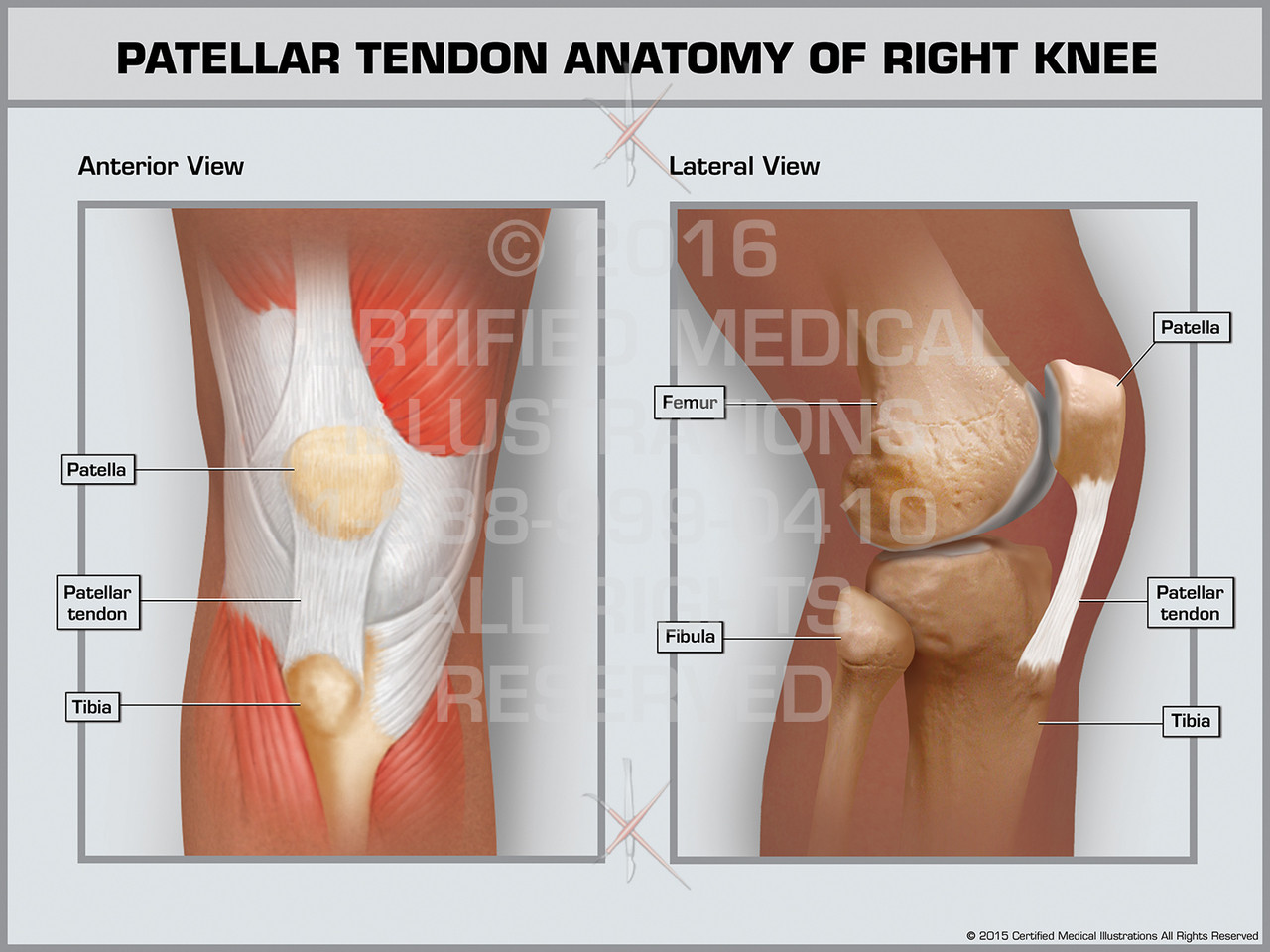
Patellar Tendon Anatomy of Right Knee from cdn10.bigcommerce.com Jun 18, 2018 · the upper leg is often called the thigh. The posterior upper leg muscles provide your knees with mobility (extension, flexion and rotation) and strength.they work closely with your quadriceps muscles at the front of your thigh, your gluteal muscles, and your calf muscles to ensure proper movement of your leg and hip. Rectus femoris, vastus lateralis, vastus medialis, and vastus intermedius. Also called the thigh bone, this is the longest bone in the body.it. There are three sets of longissimus muscles: In a complete or serious rupture the tendon of plantaris or another vestigial muscle is harvested and wrapped around the achilles tendon, increasing the strength of the repaired tendon. Quadriceps femoris muscle, large fleshy muscle group covering the front and sides of the thigh. Originates from the lateral supracondylar line of the femur.
Rectus femoris, vastus lateralis, vastus medialis, and vastus intermedius.
The tendon continues its way through the foot by extending over its dorsal surface and finally inserting on the superior surface of the base of the distal phalanx of the hallux. The longissimus (red, in the image above) are located between spinalis and the iliocostalis muscles. Apr 23, 2019 · the plantaris is a small muscle with a long tendon, which can be mistaken for a nerve as it descends down the leg. Originates from the lateral supracondylar line of the femur. The muscle descends medially, condensing into a tendon that runs down the leg, between the gastrocnemius and soleus. 1) above the cervical area (longissimus capitis), 2) in the cervical area (longissimus cervicis), and 3) in the upper back or thoracic area (longissimus thoracis). The upper arm is located between the shoulder joint and elbow joint. Quadriceps femoris muscle, large fleshy muscle group covering the front and sides of the thigh. There are three sets of longissimus muscles: Jun 18, 2018 · the upper leg is often called the thigh. They originate at the ilium (upper part of the pelvis, or hipbone) and femur (thighbone), come together in a Originating below and beneath the gastrocnemius is the soleus muscle, which extends your foot when your knee is bent. During an open surgery, an incision is made in the back of the leg and the achilles tendon is stitched together.
Originating below and beneath the gastrocnemius is the soleus muscle, which extends your foot when your knee is bent. Rectus femoris, vastus lateralis, vastus medialis, and vastus intermedius. The tendon continues its way through the foot by extending over its dorsal surface and finally inserting on the superior surface of the base of the distal phalanx of the hallux. Originates from the lateral supracondylar line of the femur. Jun 18, 2018 · the upper leg is often called the thigh.
Upper Legs Muscles Anatomy stock photo 505080183 | iStock from media.istockphoto.com The tendon continues its way through the foot by extending over its dorsal surface and finally inserting on the superior surface of the base of the distal phalanx of the hallux. During an open surgery, an incision is made in the back of the leg and the achilles tendon is stitched together. It is absent in 10% of people. They originate at the ilium (upper part of the pelvis, or hipbone) and femur (thighbone), come together in a In a complete or serious rupture the tendon of plantaris or another vestigial muscle is harvested and wrapped around the achilles tendon, increasing the strength of the repaired tendon. Also called the thigh bone, this is the longest bone in the body.it. Rectus femoris, vastus lateralis, vastus medialis, and vastus intermedius. The longissimus (red, in the image above) are located between spinalis and the iliocostalis muscles.
The longissimus (red, in the image above) are located between spinalis and the iliocostalis muscles.
They originate at the ilium (upper part of the pelvis, or hipbone) and femur (thighbone), come together in a Rectus femoris, vastus lateralis, vastus medialis, and vastus intermedius. Originating below and beneath the gastrocnemius is the soleus muscle, which extends your foot when your knee is bent. During an open surgery, an incision is made in the back of the leg and the achilles tendon is stitched together. Jun 18, 2018 · the upper leg is often called the thigh. 1) above the cervical area (longissimus capitis), 2) in the cervical area (longissimus cervicis), and 3) in the upper back or thoracic area (longissimus thoracis). The upper arm is located between the shoulder joint and elbow joint. There are three sets of longissimus muscles: Quadriceps femoris muscle, large fleshy muscle group covering the front and sides of the thigh. Originates from the lateral supracondylar line of the femur. In a complete or serious rupture the tendon of plantaris or another vestigial muscle is harvested and wrapped around the achilles tendon, increasing the strength of the repaired tendon. The tendon continues its way through the foot by extending over its dorsal surface and finally inserting on the superior surface of the base of the distal phalanx of the hallux. It is absent in 10% of people.
It is absent in 10% of people. The muscle descends medially, condensing into a tendon that runs down the leg, between the gastrocnemius and soleus. Also called the thigh bone, this is the longest bone in the body.it. It's the area that runs from the hip to the knee in each leg. Rectus femoris, vastus lateralis, vastus medialis, and vastus intermedius.
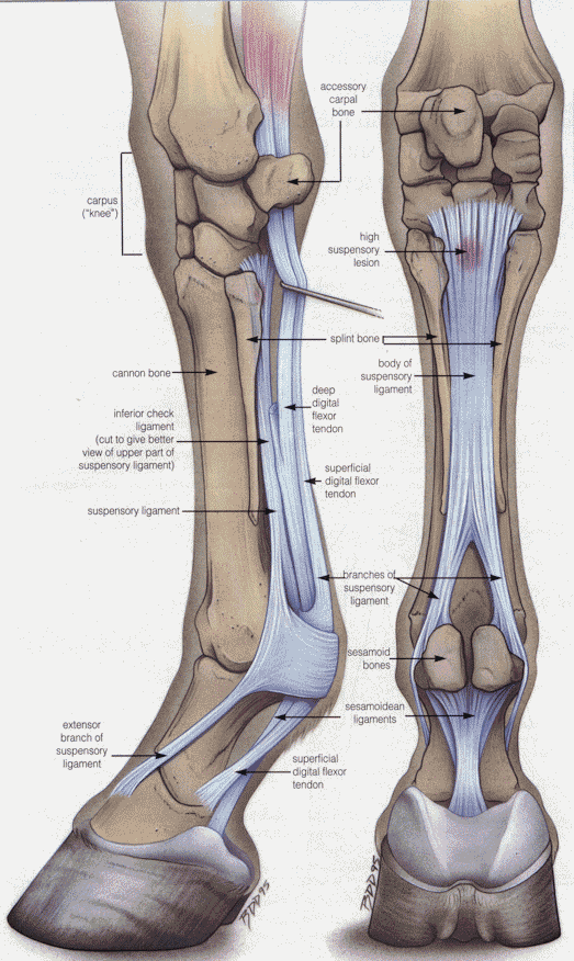
Tendon and Ligament Injuries in the Horse and Recovery ... from www.secondvet.com The muscle descends medially, condensing into a tendon that runs down the leg, between the gastrocnemius and soleus. Also called the thigh bone, this is the longest bone in the body.it. The upper arm is located between the shoulder joint and elbow joint. Originating below and beneath the gastrocnemius is the soleus muscle, which extends your foot when your knee is bent. In a complete or serious rupture the tendon of plantaris or another vestigial muscle is harvested and wrapped around the achilles tendon, increasing the strength of the repaired tendon. Quadriceps femoris muscle, large fleshy muscle group covering the front and sides of the thigh. It is absent in 10% of people. The longissimus (red, in the image above) are located between spinalis and the iliocostalis muscles.
Apr 23, 2019 · the plantaris is a small muscle with a long tendon, which can be mistaken for a nerve as it descends down the leg.
Apr 23, 2019 · the plantaris is a small muscle with a long tendon, which can be mistaken for a nerve as it descends down the leg. The tendon continues its way through the foot by extending over its dorsal surface and finally inserting on the superior surface of the base of the distal phalanx of the hallux. The posterior upper leg muscles provide your knees with mobility (extension, flexion and rotation) and strength.they work closely with your quadriceps muscles at the front of your thigh, your gluteal muscles, and your calf muscles to ensure proper movement of your leg and hip. It is absent in 10% of people. Originating below and beneath the gastrocnemius is the soleus muscle, which extends your foot when your knee is bent. 1) above the cervical area (longissimus capitis), 2) in the cervical area (longissimus cervicis), and 3) in the upper back or thoracic area (longissimus thoracis). The muscle descends medially, condensing into a tendon that runs down the leg, between the gastrocnemius and soleus. It's the area that runs from the hip to the knee in each leg. Rectus femoris, vastus lateralis, vastus medialis, and vastus intermedius. There are three sets of longissimus muscles: Originates from the lateral supracondylar line of the femur. Also called the thigh bone, this is the longest bone in the body.it. The longissimus (red, in the image above) are located between spinalis and the iliocostalis muscles.